Skip to content

Urgent urology symptoms What to do

Pelvic Circulation

Urology TodayPublished June 18, 20103 min read

Key points

  • The arterial blood supply to the pelvis and perineum are briefly reviewed.
  • In pelvic reconstructive surgery, the pertinent branches of the femoral artery are the external pudendal artery to the labial fibrofatty pad and the inferior epigastric artery, which supplies the rectus abdominus muscle.
  • The anterior trunk has seven branches: superior vesical, middle rectal, inferior vesical, uterine, internal pudendal, obturator, and inferior gluteal arteries (1).
  • The obturator canal is an opening in the membrane overlying the obturator internus through which the obturator vessels and nerve pass.

The arterial blood supply to the pelvis and perineum are briefly reviewed. Venous drainage for the most part parallels the arterial vessels. The aorta bifurcates at the level of the fourth lumbar vertebrae into the common iliac arteries (Fig. 8). The arteries run anterior and lateral to the veins at this level (1).

The common iliacs divide into the external and internal iliac arteries at the level of the sacroiliac joint. The external iliac runs along the medial aspect of the iliopsoas and exits the pelvis posterior to the inguinal ligament as the femoral artery.

In pelvic reconstructive surgery, the pertinent branches of the femoral artery are the external pudendal artery to the labial fibrofatty pad and the inferior epigastric artery, which supplies the rectus abdominus muscle. These relationships are important during rotational flap techniques.

The internal iliac artery divides into an anterior and posterior trunk. The posterior trunk gives off the superior gluteal, ascending lumbar, and lateral sacral arteries.

The anterior trunk has seven branches: superior vesical, middle rectal, inferior vesical, uterine, internal pudendal, obturator, and inferior gluteal arteries (1). The course and relationships of several of the branches are highlighted as they pertain to pelvic floor surgery.
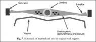
Schematic of urethral and anterior vaginal wall support

Fig. 7. Schematic of urethral and anterior vaginal wall support.

The obturator canal is an opening in the membrane overlying the obturator internus through which the obturator vessels and nerve pass.

The obturator nerve arises from the L2 through S1 nerve roots and exits the pelvis through the obturator canal, where it divides into anterior and posterior divisions to supply the muscles of the adductor compartment.

The obturator canal is nearly 5 cm superolateral to the midpoint of the ischiopubic ramus. This relationship should be kept in mind when passing a device around the ischiopubic ramus for a transobturator procedure (5).
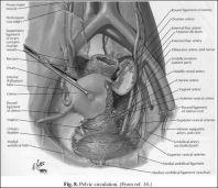
Pelvic circulation

Fig. 8. Pelvic circulation. (From ref. 16.)

The internal pudendal vessels and the pudendal nerve (S2-S4) exit the pelvis through the greater sciatic foramen, wrapping around the ischial spine and SSL laterally (Fig. 9).

Care must be taken to avoid these structures when placing sutures in the SSL. The pudendal nerve and vessels then pass through the lesser sciatic foramen and course alongside the lateral aspect of the ischiorectal fossa to enter the pudendal canal (Alcock’s canal) in the posterior perineum. The pudendal nerve branches into the inferior rectal, perineal, and dorsal (clitoral) nerves (6).
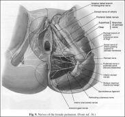
Nerves of the female perineum

Fig. 9. Nerves of the female perineum. (From ref. 16.)

Understanding lymphatic drainage is mainly significant in treating pelvic malignancy but can be relevant in inflammatory states. The pattern of lymphatic drainage parallels the venous circulation.

The vulvar lymphatic vessels, including the distal urethra and lower third of the vagina, drain into the inguinal lymph nodes. The bladder, proximal urethra, uterus, and upper two-thirds of the vagina drain into the pelvic lymph nodes (obturator, internal, and external iliac).

The ovarian lymphatics follow the gonadal vessels and drain into the para-aortic nodes (3).

—-
Melissa Fischer, MD, Priya Padmanabhan, MD, and Nirit Rosenblum, MD

—-

{embed=“ref/1”}

The content on this site is for information only and is not a substitute for professional medical advice, diagnosis or treatment. If you cannot pass urine, have a fever with back or side pain, or sudden severe pain in the testicle, call 911 or go to an emergency department.

Evaluation of Urinary Incontinence

Fluid Intake/Voiding Diary

Patient histories regarding frequency and severity of urinary symptoms are often inaccurate. A more reliable indicator of symptoms is a fluid intake/voiding diary, which should be distributed to patients. They would be instructed to document in it the amount and type of fluid consumed and the volume and time of voiding and leakage and to

5 min read

Evaluation of Urinary Incontinence

Office Evaluation of Urinary Incontinence

Introduction The goals of the basic evaluation are to confirm urinary incontinence (UI) and to identify factors that may be contributing to or resulting from UI. The general evaluation of UI should include a history, fluid intake/voiding diary, physical examination, urinalysis, urine culture, and measurement of postvoid residual urine. For selected patients, a blood test

3 min read

Anatomy of Pelvic Support

Perineum

The anatomic boundaries of the perineum are the pubis, thighs, and buttocks and can be divided into an anterior urogenital and a posterior anal triangle by drawing a line between the ischial tuberosities (Fig. 3) (7). The perineal body is a tendinous structure in the midline of the perineum between the anus and the vaginal

4 min read

Urinary symptoms?

Weak stream, urgency, up at night? Score it in two minutes.

Seven questions from the International Prostate Symptom Score show whether symptoms are mild, moderate or severe, and when to see a doctor.

Take the symptom score

When to get help

Most urology problems can wait for an appointment. These can’t.

Call 911 or go to an emergency department if you have any of these:

  • You cannot pass urine at allAcute urinary retention: a painful, swollen bladder. It needs a catheter, often within hours.
  • Fever or chills with back or side painA blocked, infected kidney (for example behind a stone) can turn into sepsis quickly.
  • Sudden, severe pain in a testiclePossible testicular torsion. The testicle can often be saved only within about 6 hours.
  • An erection lasting over 4 hoursPriapism. Without treatment it can cause permanent damage.
Call 911

Outside the US, call your local emergency number.

See a doctor within days

  1. 01Blood in your urineEven once, and even without pain. Usually harmless, but it is the most common first sign of bladder and kidney cancer.
  2. 02Burning, frequent or cloudy urineTypical of a urinary tract infection, which usually clears with treatment.
  3. 03A lump or swelling in a testicleMost lumps are benign; testicular cancer is very treatable when found early.

General guidance only. If you are unsure how urgent a symptom is, call your doctor or a nurse advice line.