
A. RENAL TRANSPLANTATION
Urinary stones associated with renal transplantation are rare. Perirenal nerves are severed at the time of renal harvesting. Classic renal colic is not found in these patients.
The patients usually are admitted with the presumptive diagnosis of graft rejection. Only after appropriate radiographic and ultrasonic evaluation is the correct diagnosis made (Figure 16–9).
B. PREGNANCY
Renal colic is the most common nonobstetric cause of acute abdominal pain during pregnancy (Figure 16–10).
Despite marked hypercalciuria associated with pregnancy, calculi are relatively rare, with an incidence approximating 1:1500 pregnancies. Women with known urinary stone disease do not have an increased risk of stones during pregnancy. The increased filtered load of calcium, uric acid, and sodium from the 25–50% increase in glomerular filtration rate associated with pregnancy has been thought to be a responsible factor in stone development.
The fetus demands special considerations regarding the potential dangers of radiation exposure (especially during the 1st trimester), medications, anesthesia, and surgical intervention. About 90% of symptomatic calculi present during the 2nd and 3rd trimesters. Initial investigations can be undertaken with renal ultrasonography and limited abdominal x-rays with appropriate shielding. Treatment requires balancing the safety of the fetus with the health of the mother. Temporizing measures to relieve upper-tract obstruction with a double-J ureteral stent or a percutaneous nephrostomy tube can be performed under local anesthesia.
{embed=“ref/stat-menu-stone-disease”}
Treatment usually can be delayed until after delivery.
C. DYSMORPHIA
Patients with severe skeletal dysmorphia that is either congenital (spina bifida, myelomeningocele, cerebral palsy) or acquired (arthritis, traumatic spinal cord injuries) and concurrent urinary calculi represent a unique clinical situation requiring special considerations (Figure 16–11). These skeletal abnormalities may preclude appropriate positioning for ESWL or percutaneous approaches. Calculi on the concave side in a patient with severe scoliosis may eliminate percutaneous puncture access between the rib and the posterosuperior iliac spine. Retrograde manipulations may need to be performed with flexible endoscopes due to marked contractures, making conventional dorsal lithotomy positioning impossible. Many such patients have undergone supravesical urinary diversion, so that retrograde access may be limited. Risks that need to be addressed include hypercalciuria associated with immobilization, relative dehydration due to patients’ or attendants’ attempts to reduce urinary output into external collecting devices, and the potential inability to drink without assistance.
A full metabolic evaluation is even more important because these social and physical restrictions may be difficult or impossible to remedy.
D. OBESITY
Obesity is a risk factor for the development of urinary calculi. Surgical bypass procedures can cause hyperoxaluria.
Massive weight gain or loss also may precipitate stone development.
Obesity limits diagnostic and treatment options. A large pannus may limit the physical examination and misguide incisions. Ultrasound examination is hindered by the attenuation of ultrasound beams. CT, magnetic resonance imaging, fluoroscopy tables, and lithotripters all have weight limitations, and patients weighing >300 lb may be unsuited for diagnosis and treatment with these resources. Standard lithotripters have focal lengths <15 cm between the energy source and the F2 target, frequently making treatment of obese patients impossible. A large anterior pannus limits prone positioning on lithotripters. Standard Amplatz nephrostomy sheaths may not be long enough to enter the collecting system. Such sheaths may need to be advanced well below the skin. A preplaced heavy suture eases removal of such sheaths.
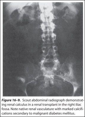
Figure 16–9. Scout abdominal radiograph demonstrating renal calculus in a renal transplant in the right iliac fossa. Note native renal vasculature with marked calcifications secondary to malignant diabetes mellitus.
“The Patch is birth control that works with my busy life.”
– ORTHO EVRA® is a contraceptive patch used for the prevention of pregnancy.
– ORTHO EVRA® is available by prescription only.
– ORTHO EVRA® does not protect against HIV (AIDS) or other sexually transmitted diseases.
– ORTHO EVRA® should not be used if you smoke cigarettes and are over 35 years old.
The ORTHO EVRA patch is used as follows: it is applied on the first day of your menstrual cycle or the first Sunday following that day, whichever is preferred. From this time on, that day is considered “patch change day.” When patch change day rolls around the following week, the first patch is removed and replaced with another. This happens once more. On the fourth patch change day the patch is removed and left off for one week, until the next patch change day when the cycle begins again.
Risks of anesthesia are increased and special high-pressure respirators may be required if patients are placed in a prone position for a percutaneous procedure. Careful positioning for open procedures helps to reduce the likelihood of crush injuries and associated rhabdomyolysis. These patients are at increased risk of anesthetic complications.
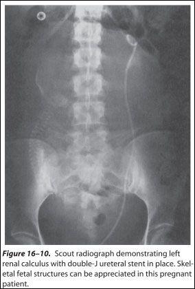
Figure 16–10. Scout radiograph demonstrating left renal calculus with double-J ureteral stent in place.
Skeletal fetal structures can be appreciated in this pregnant patient.
Postoperative prophylaxis for thromboembolic complications should be considered.
{pagebreak}
E. MEDULLARY SPONGE KIDNEY
Medullary sponge kidney is a common condition characterized by tubular ectasia associated with parenchymal cysts and clefts that predispose to nephrolithiasis in 50% of affected patients. It is most often an asymptomatic condition; however it may present with renal colic, hematuria, or urinary tract infection. It is a radiographic diagnosis.
The condition can involve select papillae or, more frequently, can be global. A full metabolic evaluation helps direct appropriate medical therapy.
F. RENAL TUBULAR ACIDOSIS
There are 3 main types of renal tubular acidosis: types I, II, and IV. Type I is associated with renal calculi.
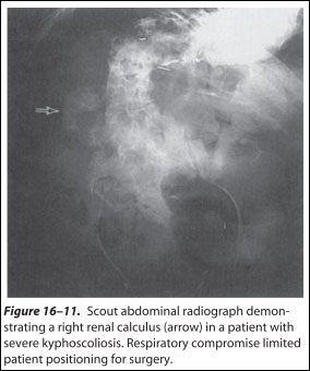
Figure 16–11. Scout abdominal radiograph demonstrating a right renal calculus (arrow) in a patient with severe kyphoscoliosis. Respiratory compromise limited patient positioning for surgery.
Patients with type I renal tubular acidosis present with persistent acidemia with a low serum bicarbonate value unexplained by hyperventilation or known renal failure. The diagnosis should be suspected in those with a known family history, severe hypocitraturia, nephrocalcinosis, medullary sponge kidney, or a fasting urine pH >6 in the absence of infection. Patients usually present with nephrolithiasis (calcium phosphate), nephrocalcinosis, or osteomalacia (or a combination). This disease can be acquired as an adult or inherited with an autosomal dominant pattern.
The diagnosis is confirmed by assessing the patient’s response to an acid load. This is frequently produced by a rapid oral ammonium chloride load (0.1 g/kg over 1 hour). The dose can be given before bedtime in the evening; the patient is instructed to fast until a second morning voided urine sample and a serum bicarbonate level are obtained.
A normal person responds by eliminating the acid load in the urine, resulting in a urinary pH <5.3. Those who do not respond in this fashion can be said to have type I renal tubular acidosis.
{embed=“ref/stat-menu-stone-disease”}
Additionally, the diagnosis should be challenged in those with normal citrate values. Treatment is centered on base replacement with potassium citrate or potassium bicarbonate solutions. Urinary citrate levels can be used to monitor effective treatment.
G. ASSOCIATED TUMORS
Squamous cell carcinoma of the upper urinary tract is uncommon but has been associated with calculi in more than 50% of cases. Chronic irritation from calculi or infection may be contributory factors. Upper-tract calculi may predispose patients to transitional cell carcinoma.
H. PEDIATRIC PATIENTS
Urinary calculi are unusual in children. A full and thorough metabolic evaluation should be undertaken. Stone analysis is particularly helpful in directing these investigations. Children born prematurely and given furosemide while in the neonatal intensive care unit are at increased risk of developing urinary stone disease. Treatment may be limited by endoscope size. Preliminary data show no change in renal growth after ESWL.
{pagebreak}
I. CALICEAL DIVERTICULA
Pyelocaliceal diverticula are cystic urine-containing eventrations of the upper tract lying within the renal parenchyma; they communicate through a narrow channel into the main collecting system (Figure 16–12). These diverticula occur in approximately 0.2–0.5% of the population and are congenital in origin; up to 40% are associated with calculi. Type I diverticula are the most common and are closely related to minor calyces. Type II have a direct communication with the renal pelvis and tend to be larger and symptomatic.
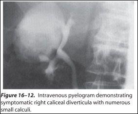
Figure 16–12. Intravenous pyelogram demonstrating symptomatic right caliceal diverticula with numerous small calculi.
Caliceal diverticuli are usually asymptomatic, but patients may complain of flank pain or recurrent urinary tract infections. Frequently many small calculi, rather than a solitary stone, are found in these obstructed cavities. When intervention was required in the past, treatment was with nephrectomy, heminephrectomy, or open surgical unroofing. Less invasive means are used today.
Communications with the collecting system are commonly pinpoint and may be difficult to locate through a retrograde approach. Retrograde access into superior pole diverticula has been successful. Surprisingly, treatment may be successful with ESWL if stone fragments are small enough to pass uneventfully. More commonly, percutaneous access and, more recently, laparoscopic means are used with success. Dilation of the caliceal neck, direct cauterization or sclerosis of the caliceal epithelium, or direct cauterization and sclerosis of the caliceal epithelium can help reduce stone recurrence rates.
{embed=“ref/stat-menu-stone-disease”}
J. RENAL MALFORMATIONS
Anatomic renal variants such as ectopic kidneys, including the horseshoe kidney, predispose to renal calculi due to impaired urinary drainage. Pain symptoms appear to be no different from those reported in patients with normally positioned kidneys. Radiographic diagnosis may be difficult due to the unexpected location of the ureters and kidneys (Figure 16–13). If calculi can be targeted with ESWL, most stone fragments pass surprisingly uneventfully.
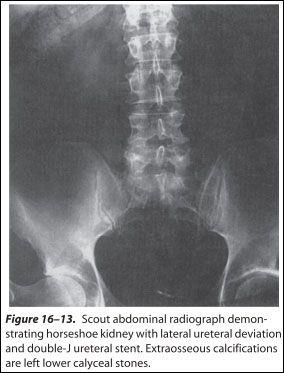
Figure 16–13. Scout abdominal radiograph demonstrating horseshoe kidney with lateral ureteral deviation and double-J ureteral stent. Extraosseous calcifications are left lower calyceal stones.
Large stone burdens should be approached percutaneously as in normally positioned kidneys. Severe outlet obstruction should be corrected with open surgery, and concurrent calculi can be removed at the same setting. Aberrant vasculature should be appreciated before percutaneous and open procedures are undertaken.
###
Marshall L. Stoller, MD
—-
REFERENCES
- Ackermann D et al: Influence of calcium content in mineral water on chemistry and crystallization conditions in urine of calcium stone formers. Eur Urol 1988;14:305.
- Allie-Hamdulay S et al: Prophylactic and therapeutic properties of a sodium citrate preparation in the management of calcium oxalate urolithiasis: Randomized, placebo-controlled trial. Urol Res 2005;33:116.
- Bilezikian JP et al: Primary hyperparathyroidism: New concepts in clinical, densitometric and biochemical features. J Intern Med 2005;257:6.
- Fellstrom B et al: Dietary habits in renal stone patients compared with healthy subjects. Br J Urol 1989;63:575.
- Gentle DL et al: Geriatric nephrolithiasis. J Urol 1997;158:2221.
- Heller HJ et al: Effect of dietary calcium on stone forming propensity. J Urol 2003;169:470.
- Langley SE, Fry CH: The influence of pH on urinary ionized [Ca2+]: